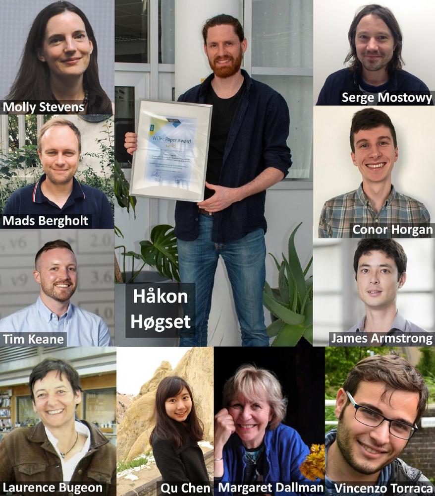
- GOLD: H. Høgset, C. C. Horgan, J. P. K. Armstrong, M. S. Bergholt, V. Torraca, Q. Chen, T. J. Keane, L. Bugeon, M. J. Dallman, S. Mostowy, M. M. Stevens (2020) In vivo biomolecular imaging of zebrafish embryos using confocal Raman spectroscopy. Nature Communications 11: 6172 www.doi.org/10.1038/s41467-020-19827-1
- SILVER: M. Yesiltas, M. Kaya, T. D. Glotch, R. Brunetto, A. Maturilli, J. Helbert, M. E. Özel (2020) Biconical reflectance, micro-Raman, and nano-FTIR spectroscopy of the Didim (H3-5) meteorite: Chemical content and molecular variations. Meteoritics & Planetary Science 55: 2404-2421 www.doi.org/10.1111/maps.13585
- BRONZE: C. Barrett, Z. Stein, J. Hernandez, R. Naraparaju, U. Schulz, L. Tetard, S. Raghavan (2021) Detrimental effects of sand ingression in jet engine ceramic coatings captured with Raman-based 3D rendering. Journal of the European Ceramic Society 41: 1664-1671 (available online 2020) www.doi.org/10.1016/j.jeurceramsoc.2020.09.050
For a list of all previous Paper Award winners, please visit www.witec.de/paper-award.
The Paper Award GOLD: Raman imaging of zebrafish embryos
Zebrafish are well-established model organisms in the life sciences and are frequently used for studying embryonic development and various diseases. Håkon Høgset from Imperial College London (ICL), UK, receives the Gold Paper Award 2021 for demonstrating the versatility of confocal Raman imaging for the biomolecular characterization of zebrafish embryos. Together with his co-workers from ICL and the London School of Hygiene & Tropical Medicine, he established that the distribution of various biomolecules such as lipids and proteins can be visualized in an embryo on different length scales. First, 3D Raman images of entire, several-millimeter-long zebrafish embryos demonstrated Raman imaging of an entire organism. Second, high-resolution Raman imaging revealed microscale features of tissue sections from dorsal muscle, tail and gut. Raman imaging was next used to detect clusters of mycobacterial infection in a zebrafish model for tuberculosis. Based on metabolic differences, Raman spectroscopy could even distinguish between infections arising from different strains. Lastly, time-lapse Raman imaging monitored molecular changes during wound response in living embryos over several hours. The authors expect that, “the ability to perform volumetric and in vivo imaging in unlabeled embryos should provide a host of new opportunities for zebrafish research that can readily complement existing fluorescence imaging techniques.”
The Paper Award SILVER: Chemical characterization of meteorites
From the chemical composition of meteorites, planetary scientists can learn a great deal about their parent bodies’ history. “Studying meteorites and their parent bodies helps us understand how our solar system formed and evolved,” says Mehmet Yesiltas from Kirklareli University, Turkey, winner of the Silver Paper Award 2021. His publication presents a detailed chemical analysis of the Didim meteorite, which he investigated together with his colleagues from research institutions in Turkey, the USA, France and Germany. The Didim meteorite (named after Didim, Turkey, where it fell in 2007) is a chondrite with a relatively rare and varied mineralogical composition, making it especially interesting. The authors investigated its chemical composition on different scales using three spectroscopic methods. Biconical reflectance spectroscopy was used for an initial large-scale assessment and revealed mainly anhydrous silicates. Raman imaging then allowed for a more precise characterization of the rock’s minerals, including feldspars, olivine and pyroxene, and their distributions on the micrometer scale. Also, aromatic hydrocarbons of different thermal metamorphic grades were shown to exist in close proximity within the meteorite. Non-destructive 3D Raman imaging showed that the carbonaceous matter was present beneath an olivine grain inside the meteorite, suggesting its extraterrestrial origin. Furthermore, nano-FTIR spectroscopy indicated that the mineralogical composition of the rock varied even on the sub-micrometer scale.
The Paper Award BRONZE: Thermochemical degradation of ceramic coatings
Jet engines are protected against their extremely high operating temperatures by thermal barrier coatings (TBCs). Ingression of molten calcium, magnesium and alumino-silicates (CMAS) into a TBC during flight causes severe damage to it and shortens the engine’s lifetime. Chance Barrett from the University of Central Florida (UCF), USA, wins the Bronze Paper Award 2021 for presenting 3D Raman imaging as a non-destructive method for analyzing the CMAS-induced degradation of TBCs, together with his co-workers from UCF and the German Aerospace Center. CMAS ingression causes a transition of the TBC to the monoclinic phase. The volume fraction of this phase therefore represents a measure of the degree of degradation and it can be quantified with Raman imaging. 3D Raman maps of TBCs visualized the degradation as a function of depth. The damage was less pronounced in the core of the columns that form the TBC than at their edges, because the gaps between the columns were more accessible. Additionally, time-dependent measurements showed that most of the damage occurred during the first hour of CMAS infiltration. The results were validated by scanning electron microscopy and energy-dispersive X-ray spectroscopy. To the authors’ knowledge, their study is the first to present a non-destructive 3D characterization of TBC degradation at high resolution. They postulate that, “This ability to quantitatively and non-destructively characterize degradation of CMAS infiltrated TBCs will accelerate development of degradation resistant coatings.”
Don’t miss your chance in the WITec Paper Award 2022
WITec invites scientists from all fields of application to participate in the Paper Award 2022 competition (www.witec.de/paper-award). Articles are eligible if they were published in 2021 in a peer-reviewed journal and feature results (at least partially) obtained with a WITec instrument. Submit your work as a PDF to papers@witec.de before January 31st, 2022. WITec is looking forward to receiving many outstanding publications again.
WITec GmbH pioneered 3D Raman imaging and correlative microscopy and continues to lead the industry with a product portfolio that offers speed, sensitivity and resolution without compromise. Raman, AFM and SNOM microscopes, combinations thereof, and WITec-developed Raman-SEM (RISE) systems can be configured for specific challenges in chemical and structural characterization through a modular hardware and software architecture with built-in capacity for expansion. Research, development and production are located at WITec headquarters in Ulm, Germany, and the WITec sales and support network has an established presence in every global region.
WITec GmbH
Lise-Meitner-Str. 6
89081 Ulm
Telefon: +49 (731) 14070-0
Telefax: +49 (731) 14070-200
https://Raman.oxinst.com
Technical Marketing & PR
Telefon: +49 (731) 140700
Fax: +49 (173) 14070200
E-Mail: eleni.kallis@witec.de
![]()

